Weeks 4&5 (Ch06)
Sensory Components of Motor Control

Associate Professor, California State University, Northridge
2025-10-05
0.2 ✋ Touch & Motor Control: Overview
- Touch provides essential feedback for achieving action goals in daily skills
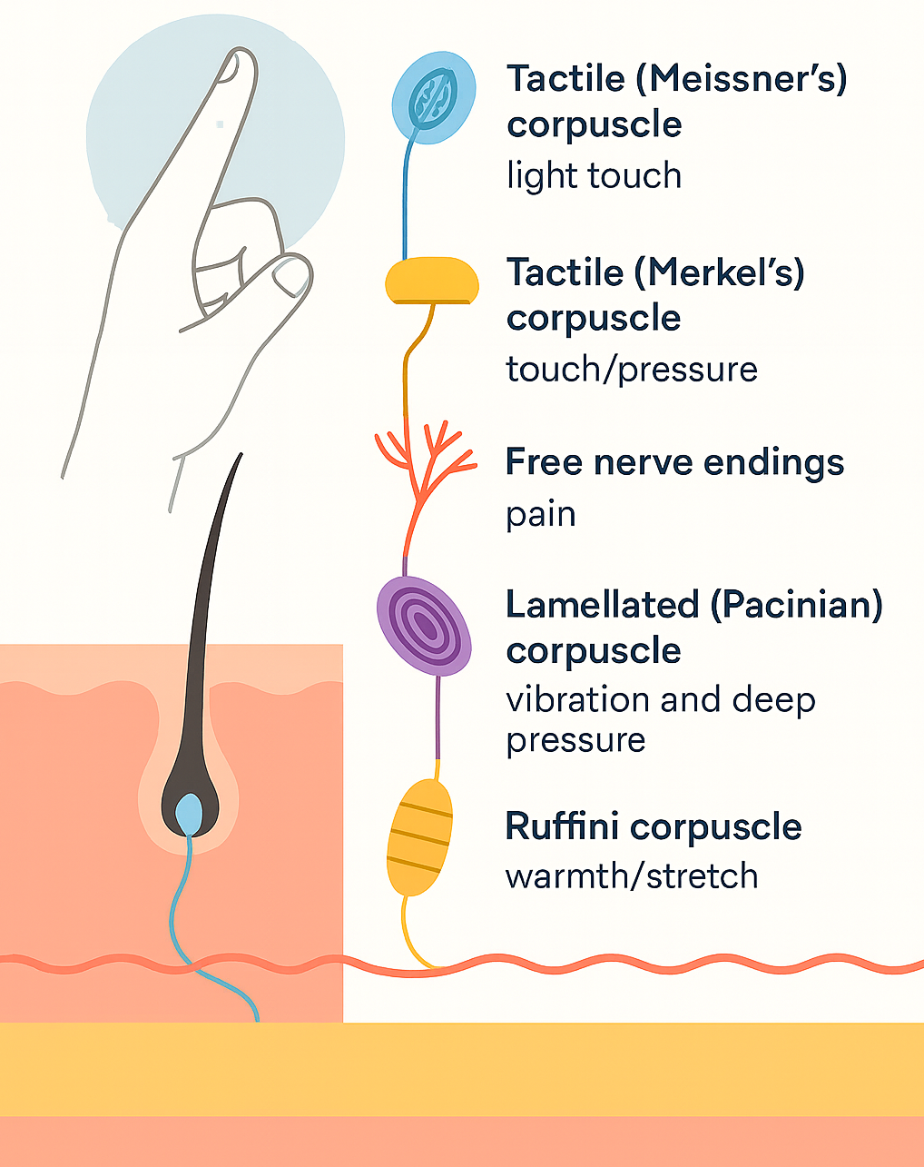
- Skin receptors (mechanoreceptors):
- Located in the dermis
- Densest in fingertips → support precision
- Signal pressure, stretch, vibration, temperature, pain
- Located in the dermis
- Critical for:
- Object manipulation (e.g., grasping, typing, playing piano)
- Interactions with people/environment (e.g., walking, sports)
- Object manipulation (e.g., grasping, typing, playing piano)
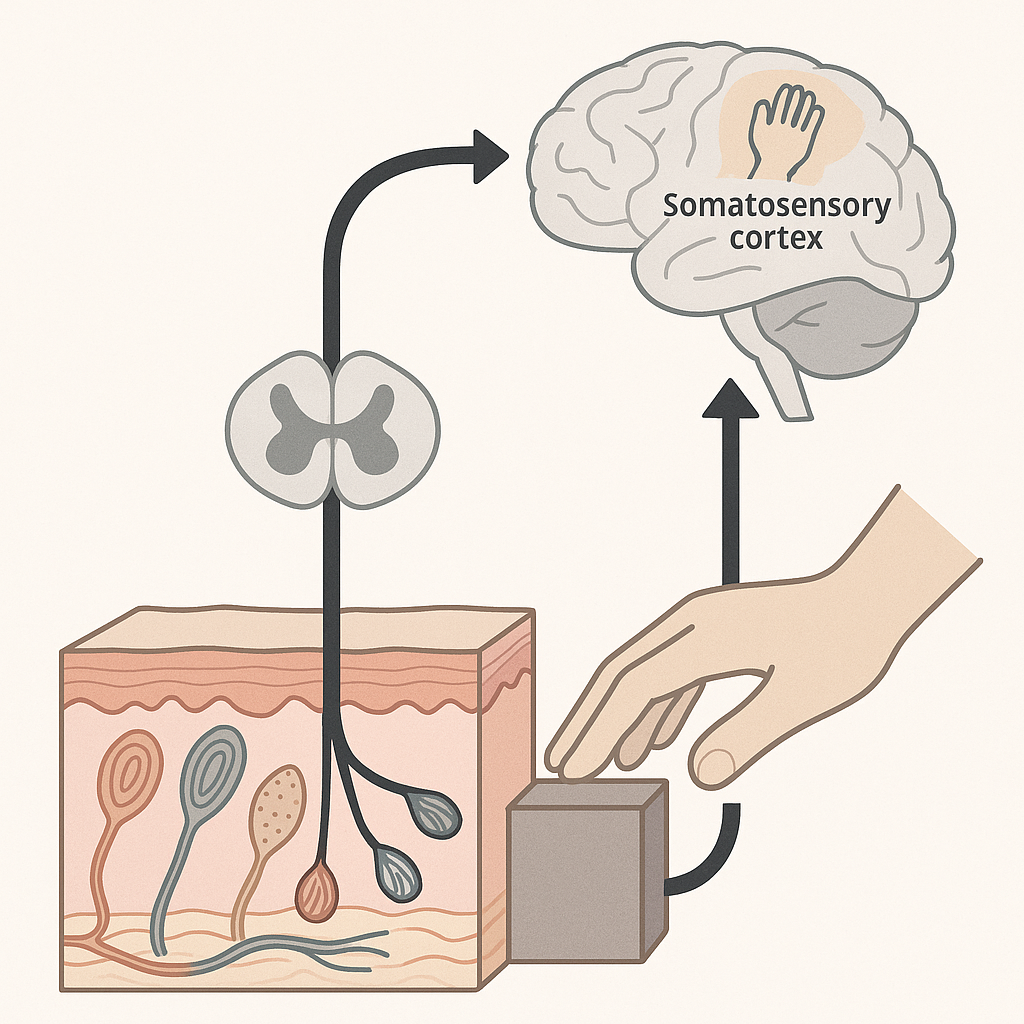
0.3 Neural Basis of Touch (at a glance)
- Mechanoreceptors in the skin transduce deformation into neural signals
- Tactile information travels via ascending somatosensory pathways
- Signals reach the somatosensory cortex, integrating with motor areas
- This feedback enables action planning, adjustment, and control
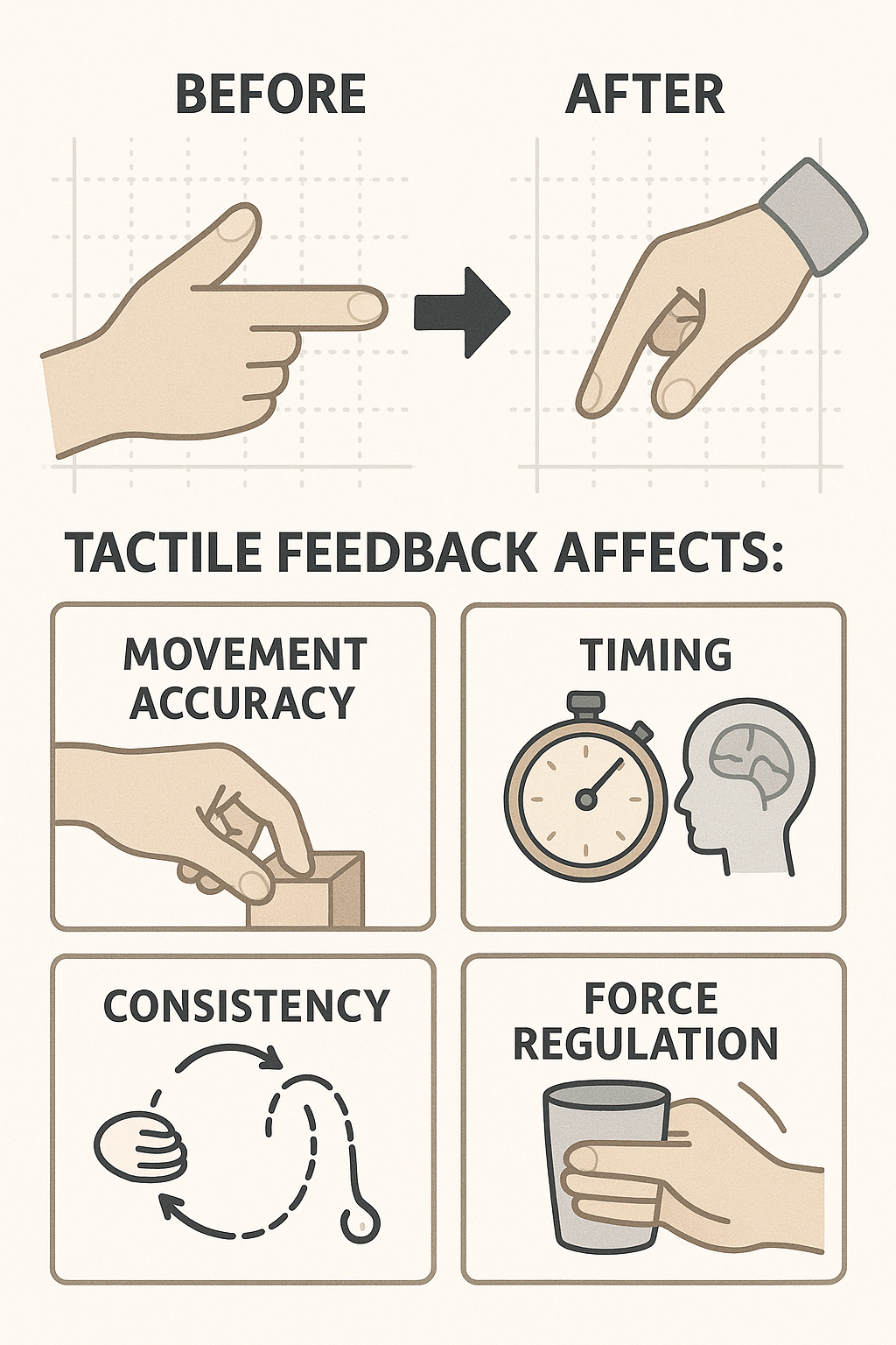
0.4 🖐️ Roles of Tactile Information in Motor Control
- Experimental approach: Compare motor performance before and after anesthetizing fingertips
- Tactile (cutaneous) feedback affects:
- ✅ Movement accuracy — especially in pointing, grasping, and fine motor skills
- 🔁 Consistency — reduces variability in repeated movements
- ⏱️ Timing — crucial for rhythmic actions and phase transitions (e.g., tapping, circle drawing)
- 🧠 Force regulation — helps scale grip force and adjust mid-movement (e.g., lifting a cup)
- ✅ Movement accuracy — especially in pointing, grasping, and fine motor skills
Tactile input supports precision, rhythm, and adaptability in everyday and skilled movements.
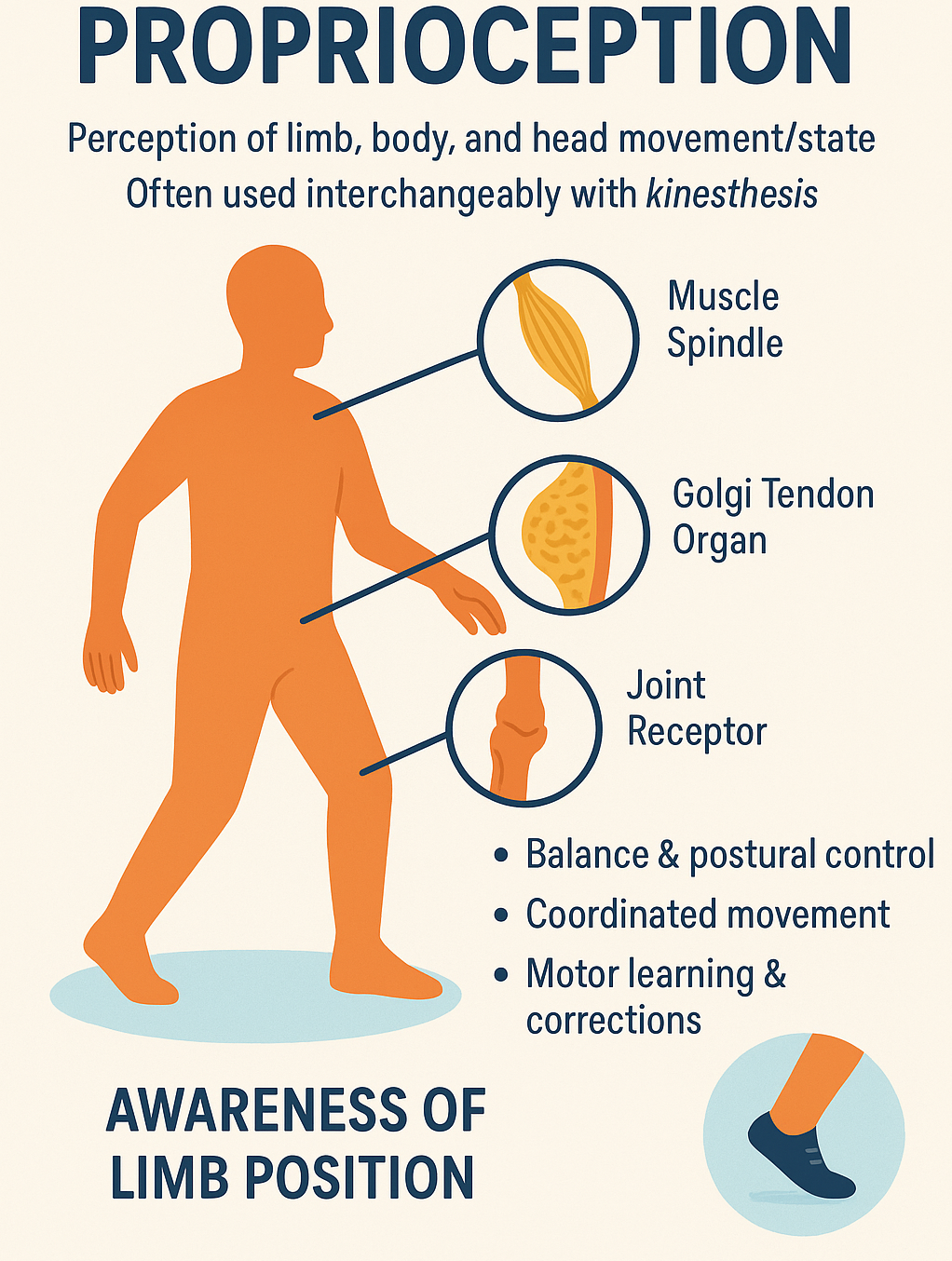
0.5 Proprioception: Definition
Proprioception is the body’s ability to sense the position, movement, and force of limbs, trunk, and head — even without visual input.
- Sometimes used interchangeably with kinesthesis
- Involves feedback from muscle spindles, Golgi tendon organs, and joint receptors
- Critical for:
- Balance and postural control
- Coordinated movement
- Motor learning and corrections
📌 Essential for executing movements like walking, reaching, and grasping — even in the dark!
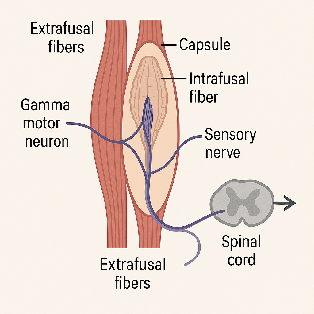
0.7 💪 Muscle Spindles (1)
- Encapsulated intrafusal fibers located within most skeletal muscles
- Arranged in parallel with force-generating extrafusal fibers
- Sensory endings (type Ia afferents) wrap around the central region → detect muscle length & velocity
- Innervated by gamma motor neurons (fusimotor system) → maintain spindle sensitivity during contraction
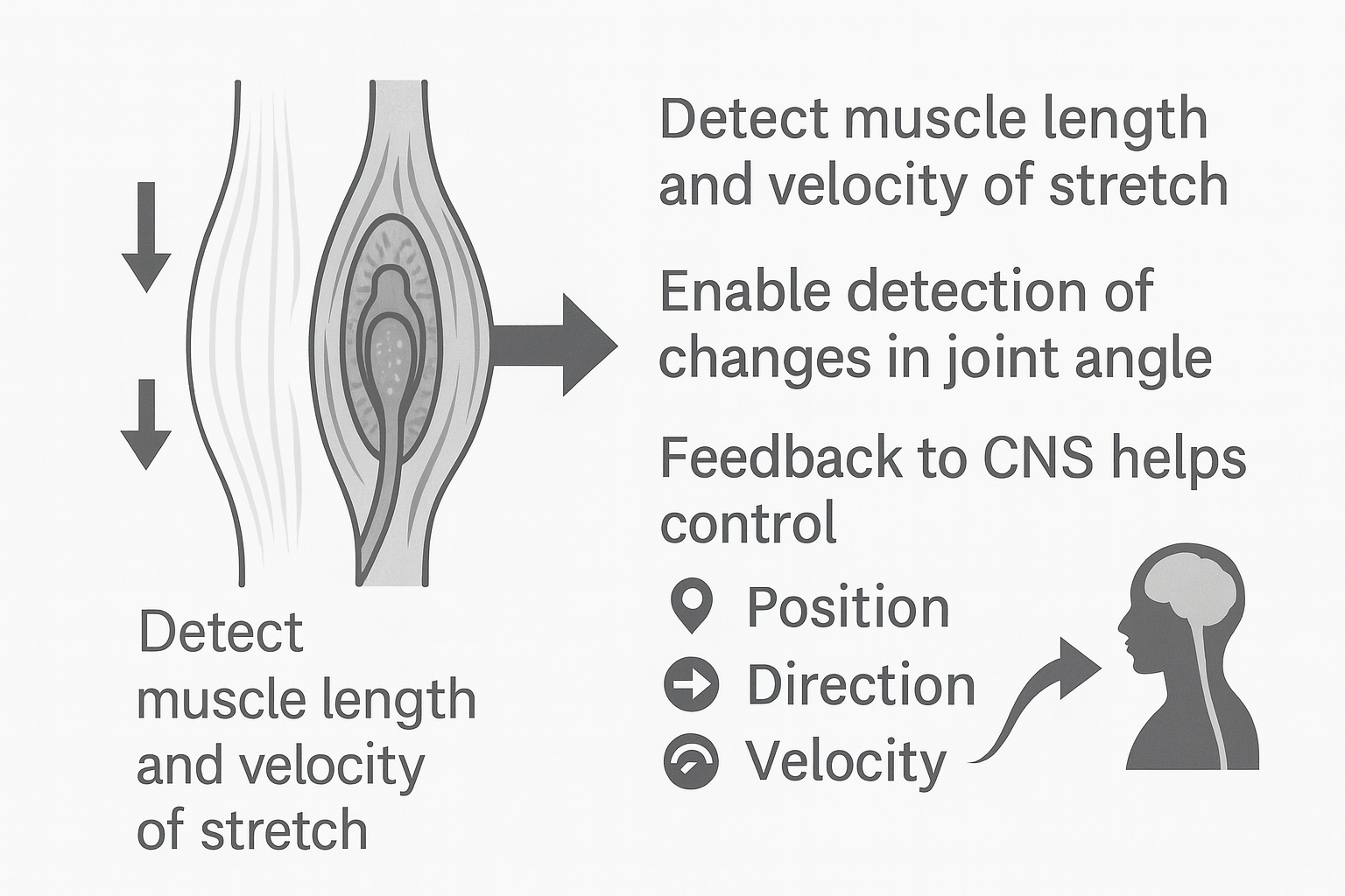
0.8 🌀 Muscle Spindles (2)
- Detect muscle length and velocity of stretch
- Provide sensory basis for joint angle changes
- Continuous feedback to CNS supports control of:
- Position (limb placement in space)
- Direction (movement trajectory)
- Velocity (speed of movement)
- Position (limb placement in space)
- Critical for both movement correction and planning
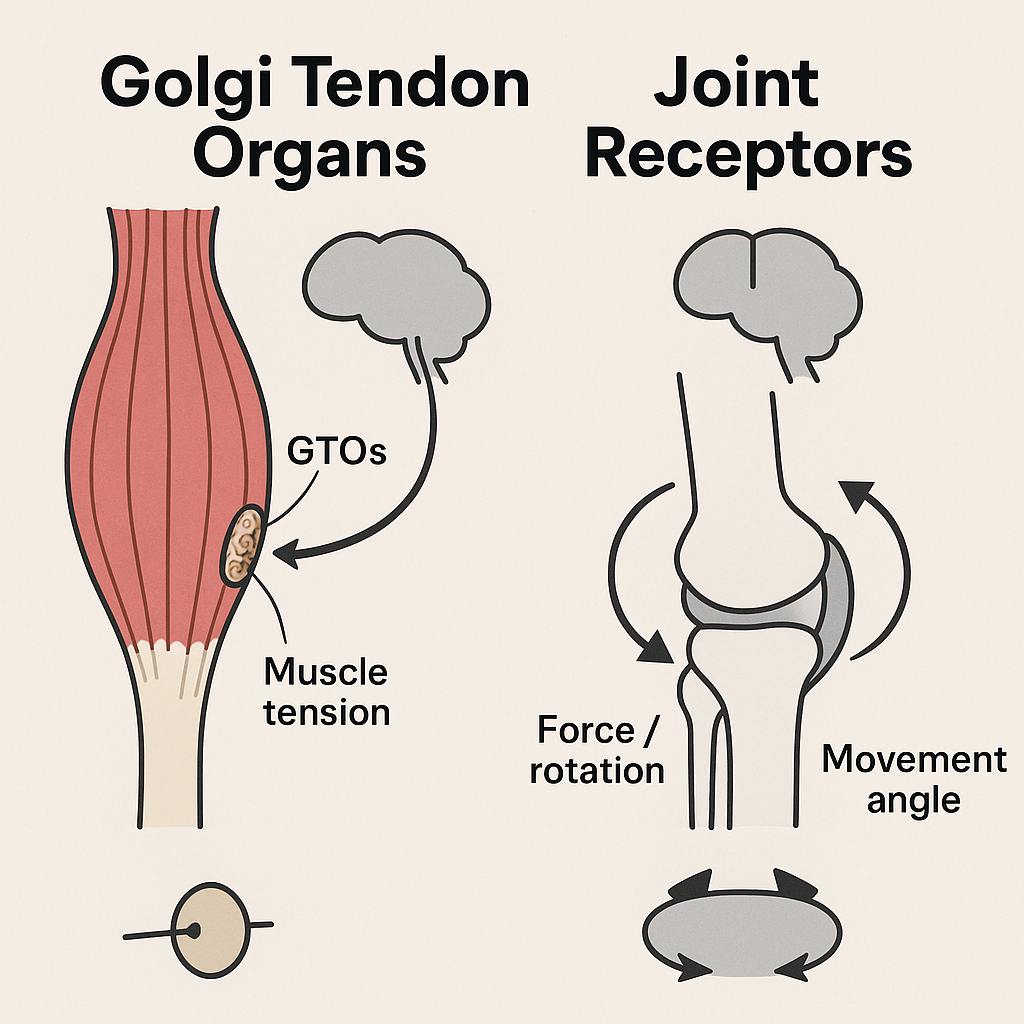
0.9 ⚖️ Golgi Tendon Organs & Joint Receptors
- Golgi Tendon Organs (GTOs):
- Located near tendon insertions in skeletal muscle
- Detect muscle tension / force (not length)
- Provide inhibitory feedback to prevent excessive force
- Located near tendon insertions in skeletal muscle
- Joint Receptors:
- Found in joint capsules and ligaments
- Detect force, rotation, and movement angle
- Especially sensitive at end ranges of motion
- Include Ruffini endings, Pacinian corpuscles, and Golgi-like receptors
- Found in joint capsules and ligaments
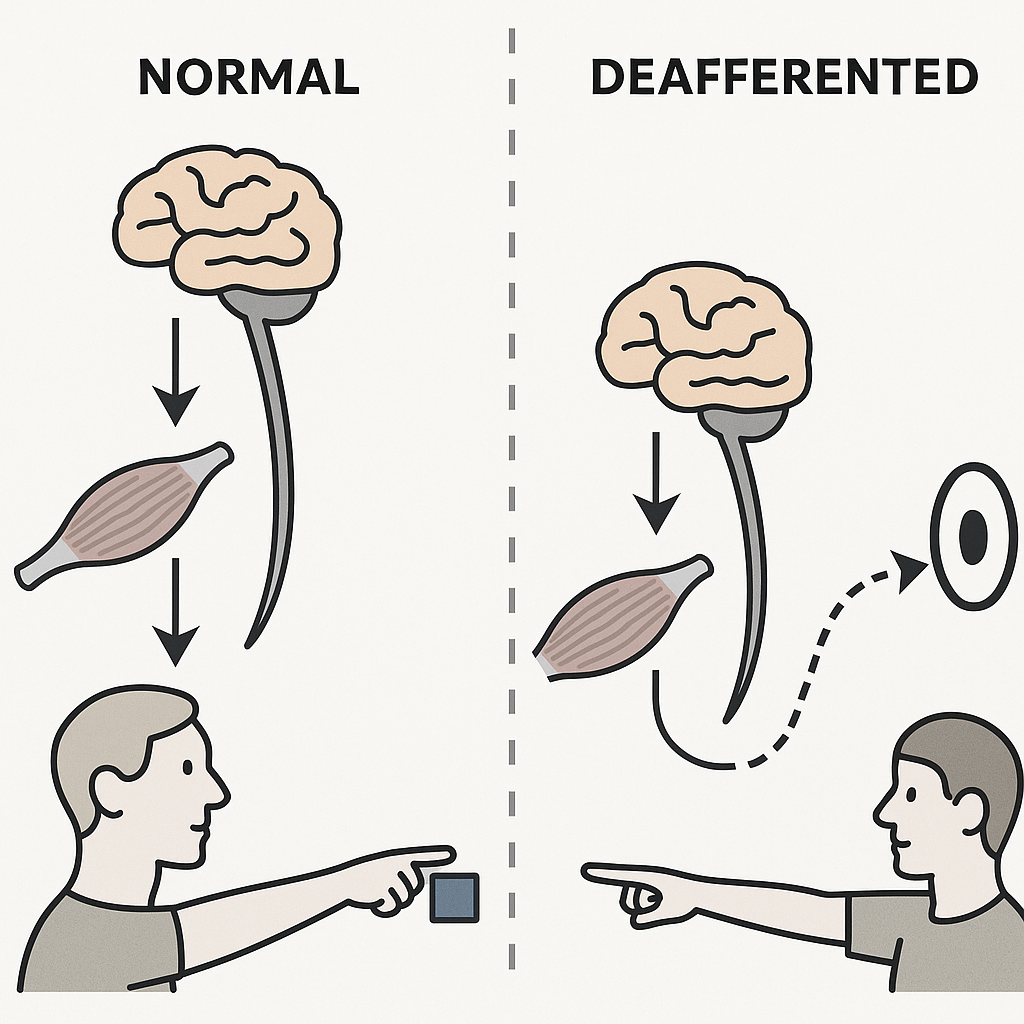
0.10 🧪 Investigating Proprioception: Deafferentation
- Surgical deafferentation
- Afferent pathways severed or removed (animal studies, rare clinical cases)
- Used to study how loss of proprioceptive input alters movement control
- Afferent pathways severed or removed (animal studies, rare clinical cases)
- Sensory neuropathy (peripheral neuropathy)
- Loss of large myelinated afferents → profound proprioceptive deficits
- Pain & temperature sensation often preserved
- Movements show spatial errors, poor smoothness, and lack of coordination
- Loss of large myelinated afferents → profound proprioceptive deficits
- Research example: Blouin et al. (1993) cited in Magill & Anderson (2017)
- Compared deafferented patient vs. healthy controls in a pointing task
- With vision: performance nearly normal
- Without vision: patient consistently undershot targets
- Compared deafferented patient vs. healthy controls in a pointing task
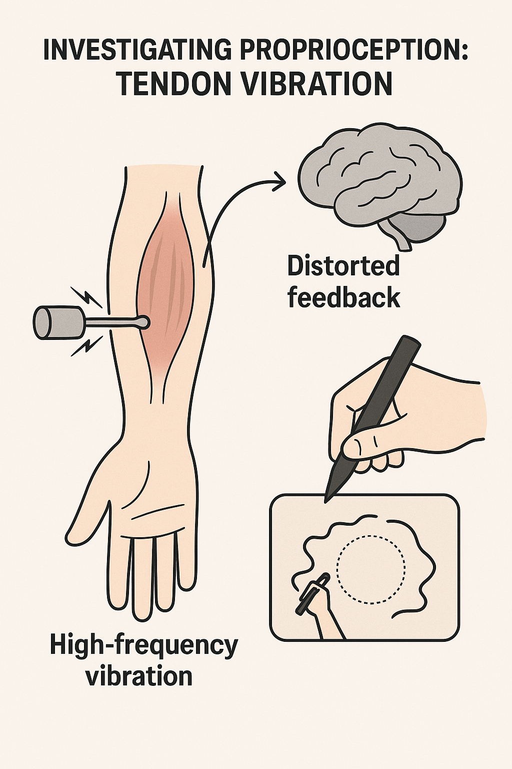
0.11 🎵 Investigating Proprioception: Tendon Vibration
Method: Apply high-frequency vibration to the tendon of an agonist muscle
Effect: Distorts proprioceptive feedback → creates illusory lengthening of the muscle
Unlike deafferentation, feedback is altered (not removed)
Used to study proprioceptive contribution to movement control & coordination
Research example: Verschueren et al. (1999) cited in Magill & Anderson (2017)
- Vibrating biceps/anterior deltoid altered arm trajectory in circle drawing
- Showed disrupted spatial accuracy and inter-limb coordination
- Vibrating biceps/anterior deltoid altered arm trajectory in circle drawing
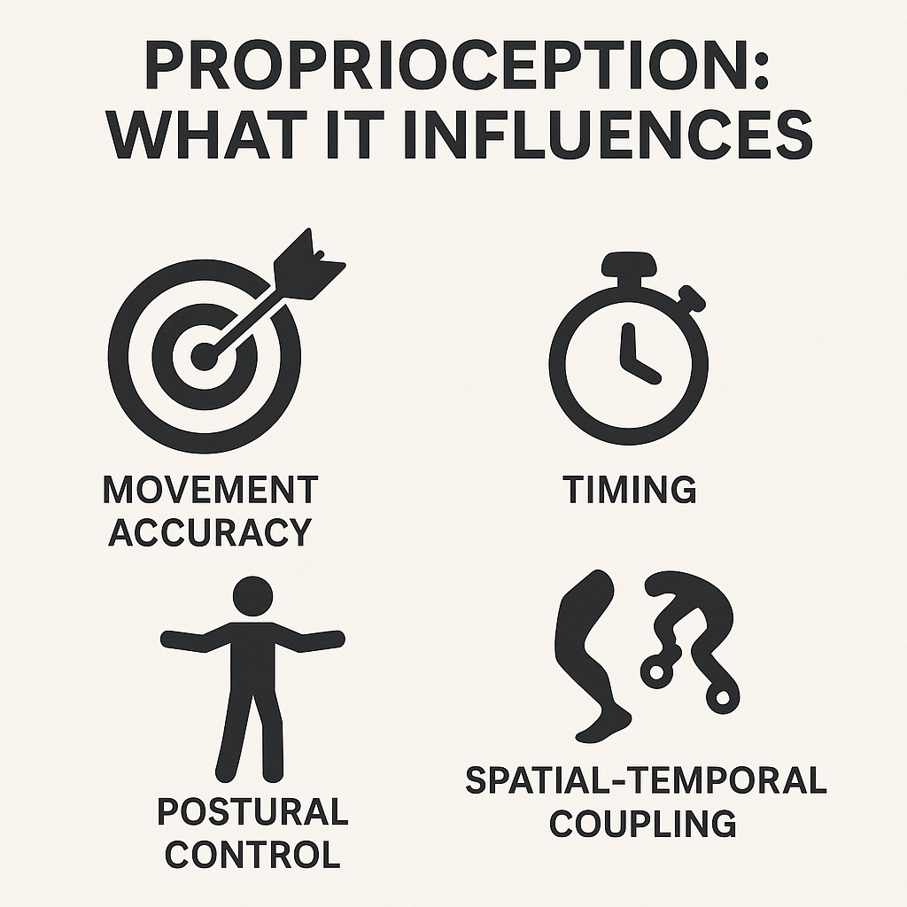
0.12 ⚙️ Proprioception: What It Influences
- Movement accuracy
- Critical for spatial & temporal precision
- Errors increase without proprioceptive feedback
- Critical for spatial & temporal precision
- Timing
- Influences onset of motor commands
- Coordinates sequencing of limb actions
- Influences onset of motor commands
- Coordination
- Supports segmental coupling within and across limbs
- Ensures smooth multi-joint movement patterns
- Supports segmental coupling within and across limbs
- Postural control
- Provides essential feedback for stabilization and balance
- Works together with vision & vestibular input
- Provides essential feedback for stabilization and balance
- Spatial–temporal coupling
- Links timing & positioning of limb segments
- Especially important in complex or bimanual tasks
- Links timing & positioning of limb segments
0.13 👁️ Vision: Neurophysiology (Overview)
- Eye → Retina → Optic nerve → Subcortical & Cortical pathways
- Retina: photoreceptors (rods & cones) transduce light → neural signals
- Parallel processing streams:
- Ventral (“vision-for-perception”) → object identification (form, color, detail)
- Dorsal (“vision-for-action”) → spatial guidance & movement control
- Ventral (“vision-for-perception”) → object identification (form, color, detail)
- Integration with motor areas enables targeting, tracking, and corrections
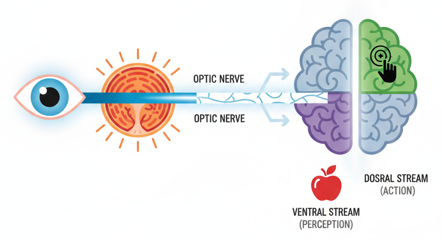
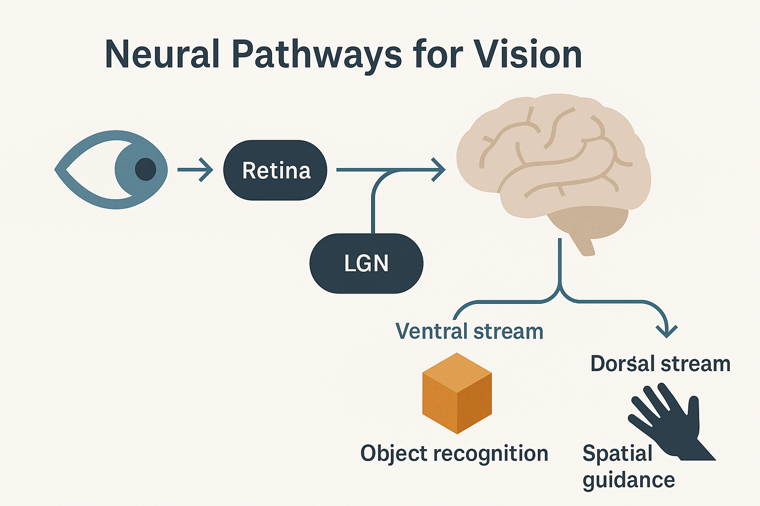
0.14 👁️ Neural Pathways for Vision (Overview)
- Light enters the retina, photoreceptors transduce light into neural signals
- Signals travel via the optic nerve and cross at the optic chiasm
- Relayed to the lateral geniculate nucleus (LGN) of the thalamus
- Projected to the primary visual cortex in the occipital lobe
- From the cortex, information diverges into two parallel streams:
- Ventral stream (vision-for-perception): object recognition and identification
- Dorsal stream (vision-for-action): spatial guidance and movement control
- Ventral stream (vision-for-perception): object recognition and identification
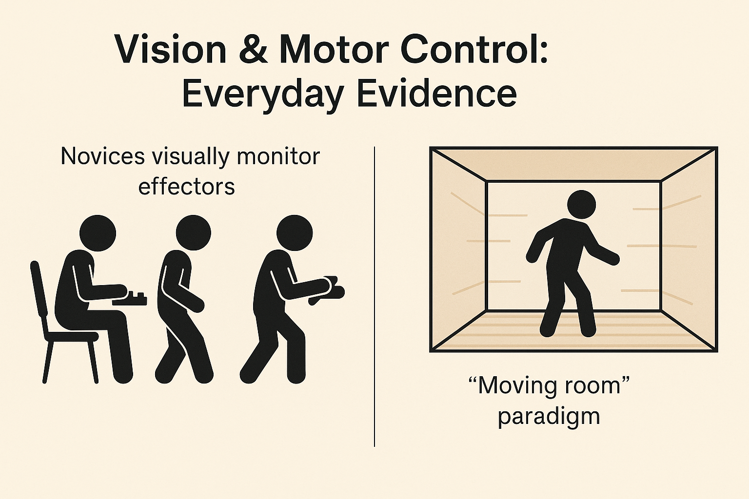
0.15 👀 Vision & Motor Control: Everyday Evidence
- Novices rely on vision to monitor effectors
- Typists looking at their fingers
- Dancers watching their feet
- New drivers visually scanning every control
- Typists looking at their fingers
- With skill development, reliance on vision decreases as tactile and proprioceptive feedback increase
- Classic “moving room” paradigm (Lee & Aronson)
- Visual cues can override proprioceptive and vestibular information
- Demonstrates visual dominance in postural control
- Visual cues can override proprioceptive and vestibular information
- Everyday life: vision provides continuous reference for balance, posture, and spatial orientation
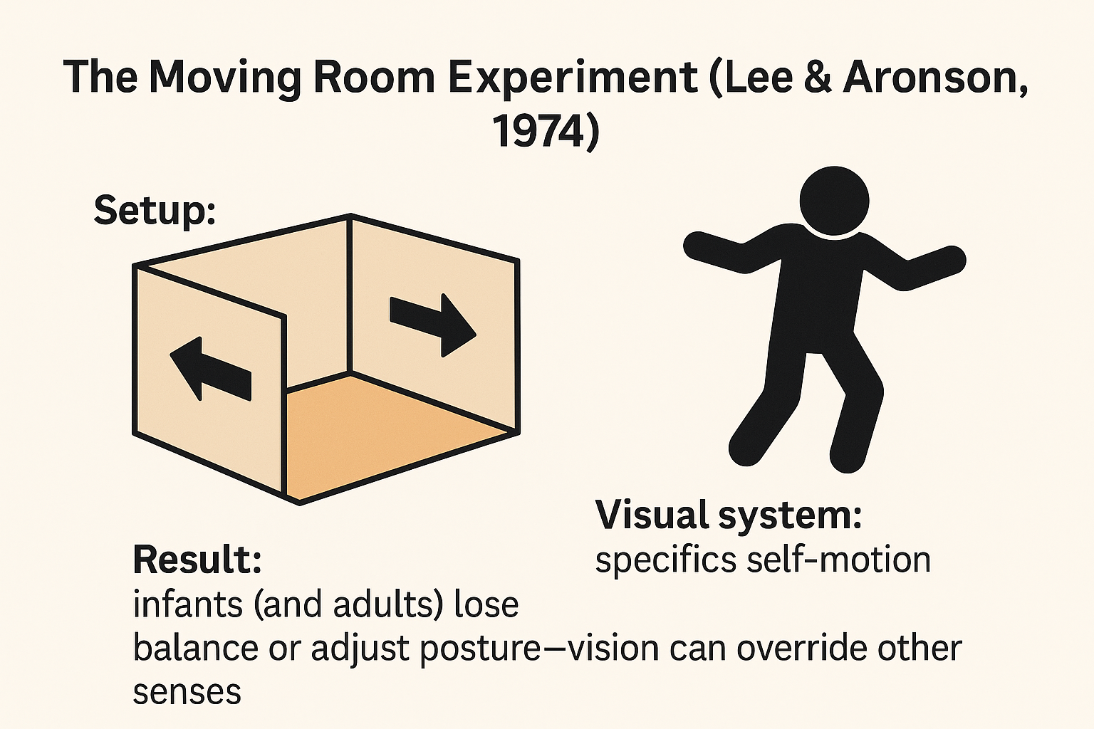
0.16 🏠 The Moving Room Experiment (Lee & Aronson, 1974 cited in Magill & Anderson (2017))
- Setup:
- Walls and ceiling of the room move back and forth
- Floor remains completely stationary
- Walls and ceiling of the room move back and forth
- Conflict of sensory information:
- Visual system: signals self-motion
- Vestibular & somatosensory systems: signal no movement
- Visual system: signals self-motion
- Findings:
- Infants and adults sway, stumble, or lose balance
- Demonstrates visual dominance—vision can override other senses in postural control
- Infants and adults sway, stumble, or lose balance
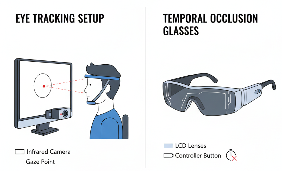
0.17 🎥 Studying Vision in Action (1): Methods
- Eye-movement recording
- Tracks where the eyes are looking and for how long
- Identifies point of gaze (foveal vision) during skill performance
- Tracks where the eyes are looking and for how long
- Temporal occlusion techniques
- Stop video at different time points to test when critical information is detected
- Use of liquid-crystal spectacles (PLATO glasses) to control viewing windows in real time
- Stop video at different time points to test when critical information is detected
- Provide insights into how vision is used for anticipation, decision-making, and movement control
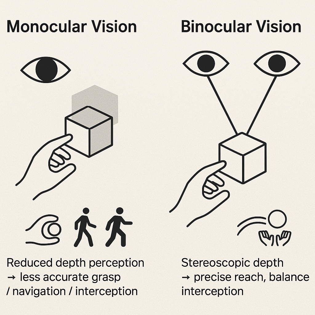
0.20 👀 Monocular vs. Binocular Vision
- Binocular vision
- Provides depth perception and 3D spatial accuracy
- Critical for:
- Reaching and grasping objects
- Walking through cluttered or uneven environments
- Intercepting moving objects (e.g., catching, hitting)
- Reaching and grasping objects
- Provides depth perception and 3D spatial accuracy
- Monocular vision
- Can support performance but with reduced accuracy and efficiency
- Leads to underestimation of distance and object size
- Errors more pronounced as distance to target increases
- Can support performance but with reduced accuracy and efficiency
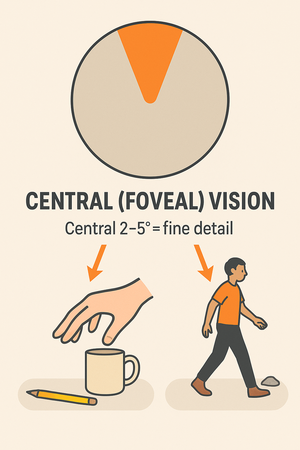
0.21 🎯 Central (Foveal) Vision
- Covers the central ~2–5° of the visual field (foveal vision)
- Responsible for detecting fine detail and specific features
- Provides critical information to support action goals:
- Reaching & grasping
- Detects object regulatory conditions (size, shape, orientation)
- Guides grip formation and movement trajectory
- Detects object regulatory conditions (size, shape, orientation)
- Locomotion
- Supplies precise path and obstacle details
- Supports accurate foot placement and navigation
- Supplies precise path and obstacle details
- Reaching & grasping
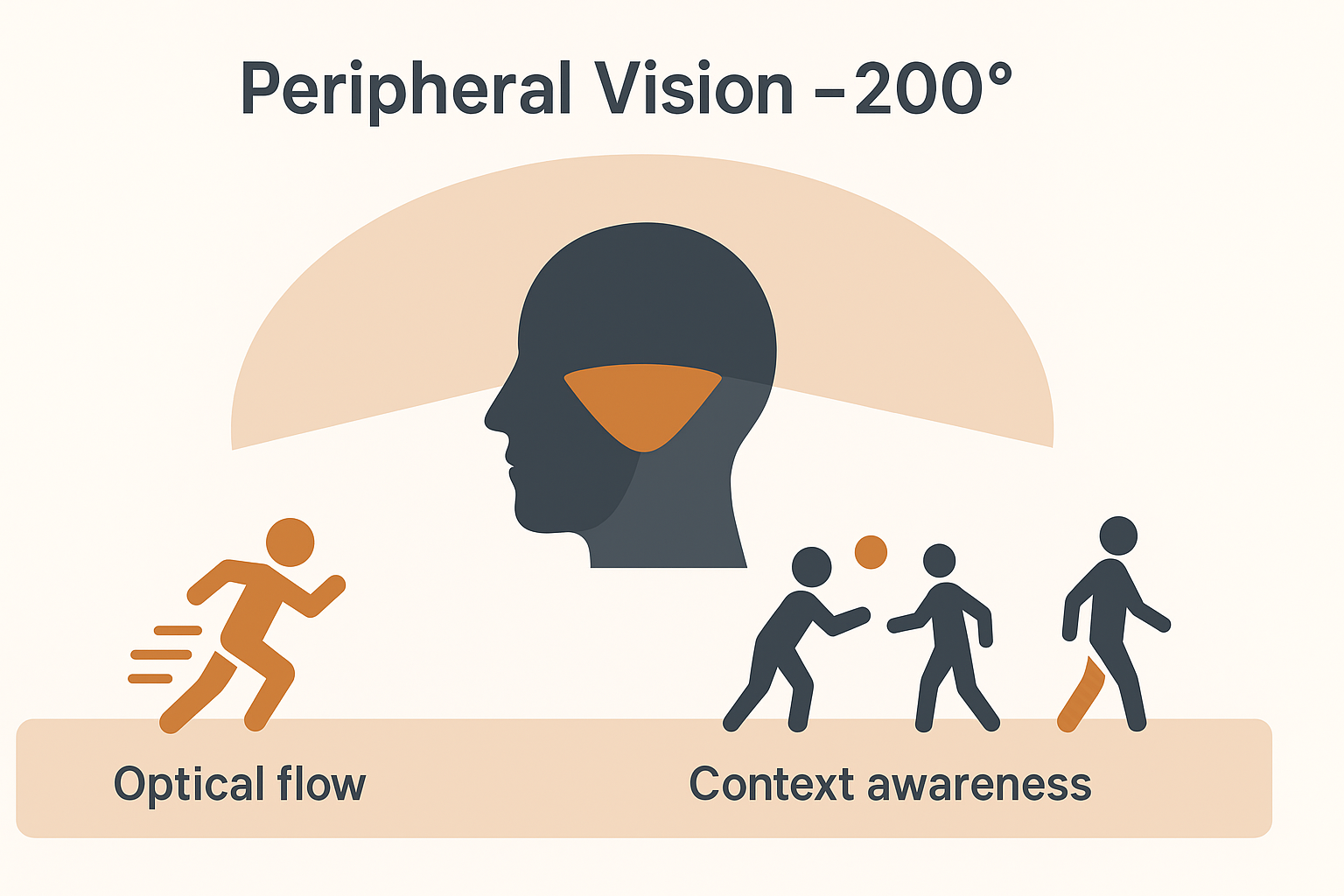
0.22 👁️ Peripheral Vision
- Extends across ~200° of the visual field
- Provides contextual information beyond the central 2–5°
- Contributes to perception of own limb movement during actions
- Essential for optical flow:
- Pattern of motion across the retina created by self-movement
- Guides posture, locomotion, and orientation in space
- Pattern of motion across the retina created by self-movement
- Supports navigation through the environment and coordination with moving objects or people
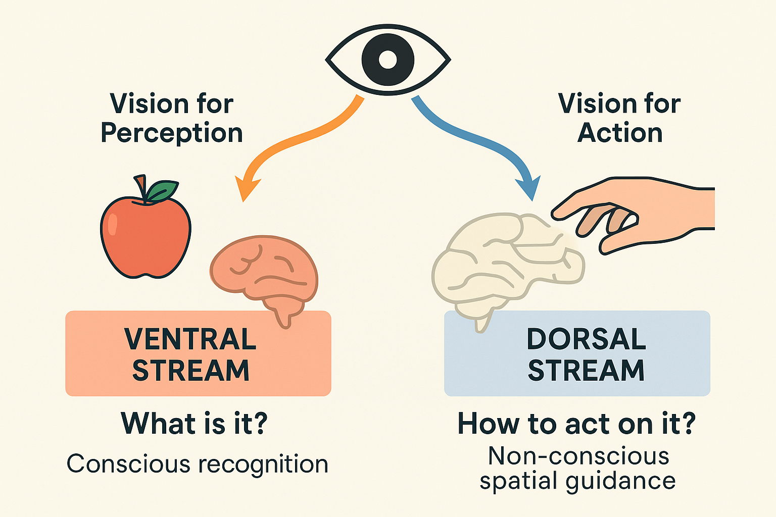
0.23 👓 Two Visual Systems
- Vision-for-Perception (Ventral stream)
- Pathway: visual cortex → temporal lobe
- Fine analysis of visual scene: form, color, features
- Supports object recognition and description
- Information is typically conscious
- Pathway: visual cortex → temporal lobe
- Vision-for-Action (Dorsal stream)
- Pathway: visual cortex → posterior parietal lobe
- Provides spatial characteristics of objects and environment
- Guides movement planning and online control
- Processing often occurs non-consciously
- Pathway: visual cortex → posterior parietal lobe
- Streams operate in parallel → perception and action are supported simultaneously
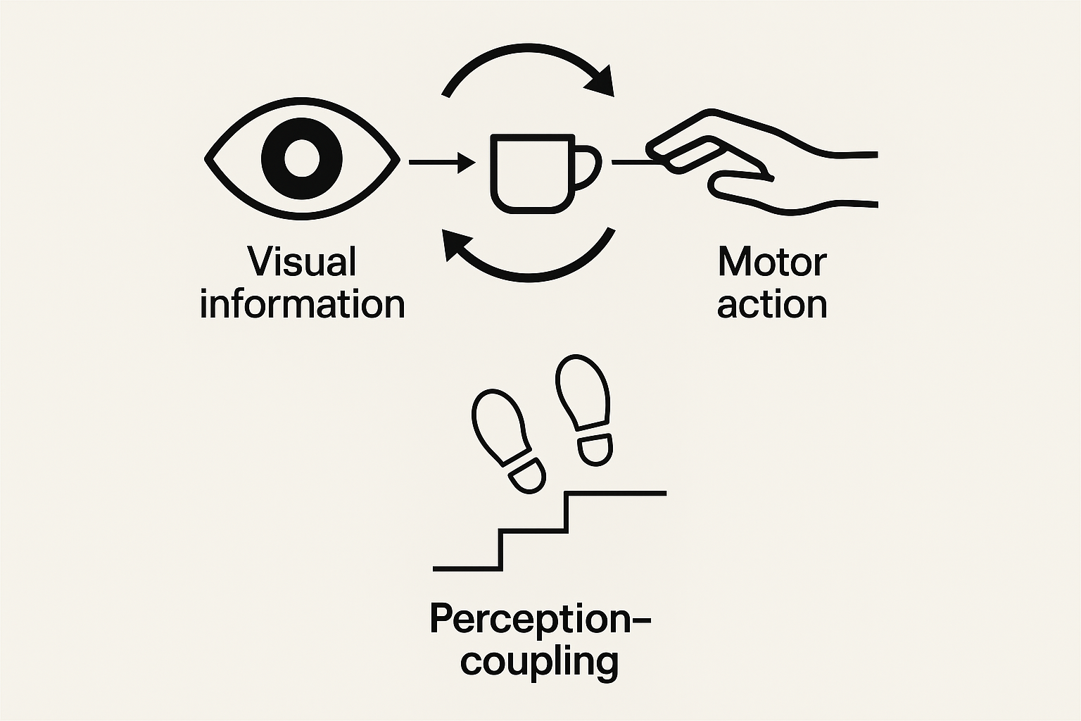
0.24 🔗 Perception–Action Coupling
- Perceptual information and motor actions are tightly connected
- Visual perception continuously informs movement parameters
- Eye–hand coordination as a classic example:
- Spatial and temporal features of gaze align with limb kinematics
- Point of gaze typically arrives at the target before the hand
- Spatial and temporal features of gaze align with limb kinematics
- Coupling ensures movements are adjusted online to match environmental demands
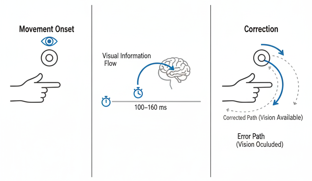
0.25 ⏱️ Online Visual Corrections: Time Required
- Experimental approach
- Compare rapid aiming when target is visible vs. occluded after movement onset
- If vision is available, corrections can be made mid-flight
- If vision is removed, errors increase
- Compare rapid aiming when target is visible vs. occluded after movement onset
- Time window for corrections
- Visual feedback requires ~100–160 ms to process
- Corrections possible only if movement duration allows this window
- Visual feedback requires ~100–160 ms to process
- Implications
- Fast, ballistic movements often too brief for corrections
- Slower or sustained movements benefit from visual feedback adjustments
- Fast, ballistic movements often too brief for corrections
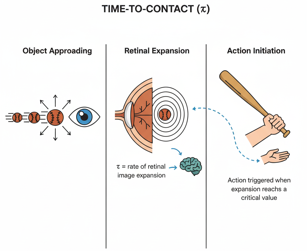
0.26 ⏳ Time-to-Contact (τ)
- In interception and avoidance tasks, vision specifies when to initiate action
- Optical variable tau (τ):
- Derived from the rate of expansion of an object’s image on the retina
- Provides a direct estimate of time remaining until contact
- Derived from the rate of expansion of an object’s image on the retina
- At a critical expansion rate, action is automatically triggered (non-conscious)
- Allows precise movement initiation in dynamic contexts:
- Catching or hitting moving objects
- Avoiding oncoming obstacles
- Timing steps or braking when approaching surfaces or vehicles
- Catching or hitting moving objects