Week 2 (Ch04)
Neuromotor Basis for Motor Control
Ovande Furtado Jr., PhD. 
Associate Professor, California State University, Northridge
2025-10-05
1.1 The Neuromotor System: An Introduction
- Start by watching this brief video on the nervous system: Nervous System Overview.
- The neuromotor system is the amazing foundation for how we control our movements, from simple to intricate tasks.
- This discussion will dive into the neurophysiological basis of actions, specifically the central nervous system’s (CNS) structure and function as it relates to motor skills.
- Understanding this process is vital for anyone planning a career in physical rehabilitation or helping people learn and relearn motor skills.
- It provides a comprehensive appreciation of human capabilities and limitations in movement.
1.2 The Neuron: Basic Building Block
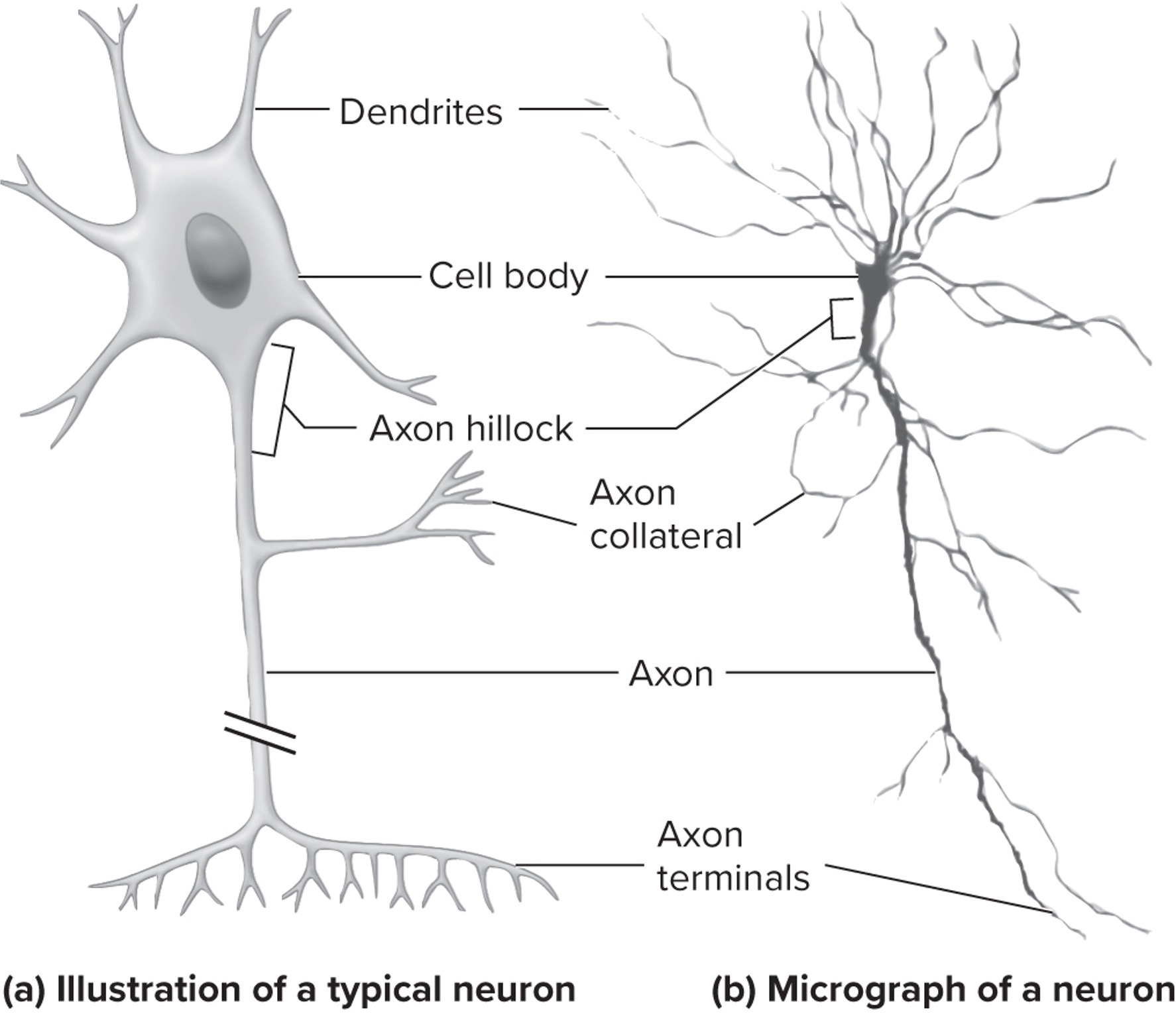
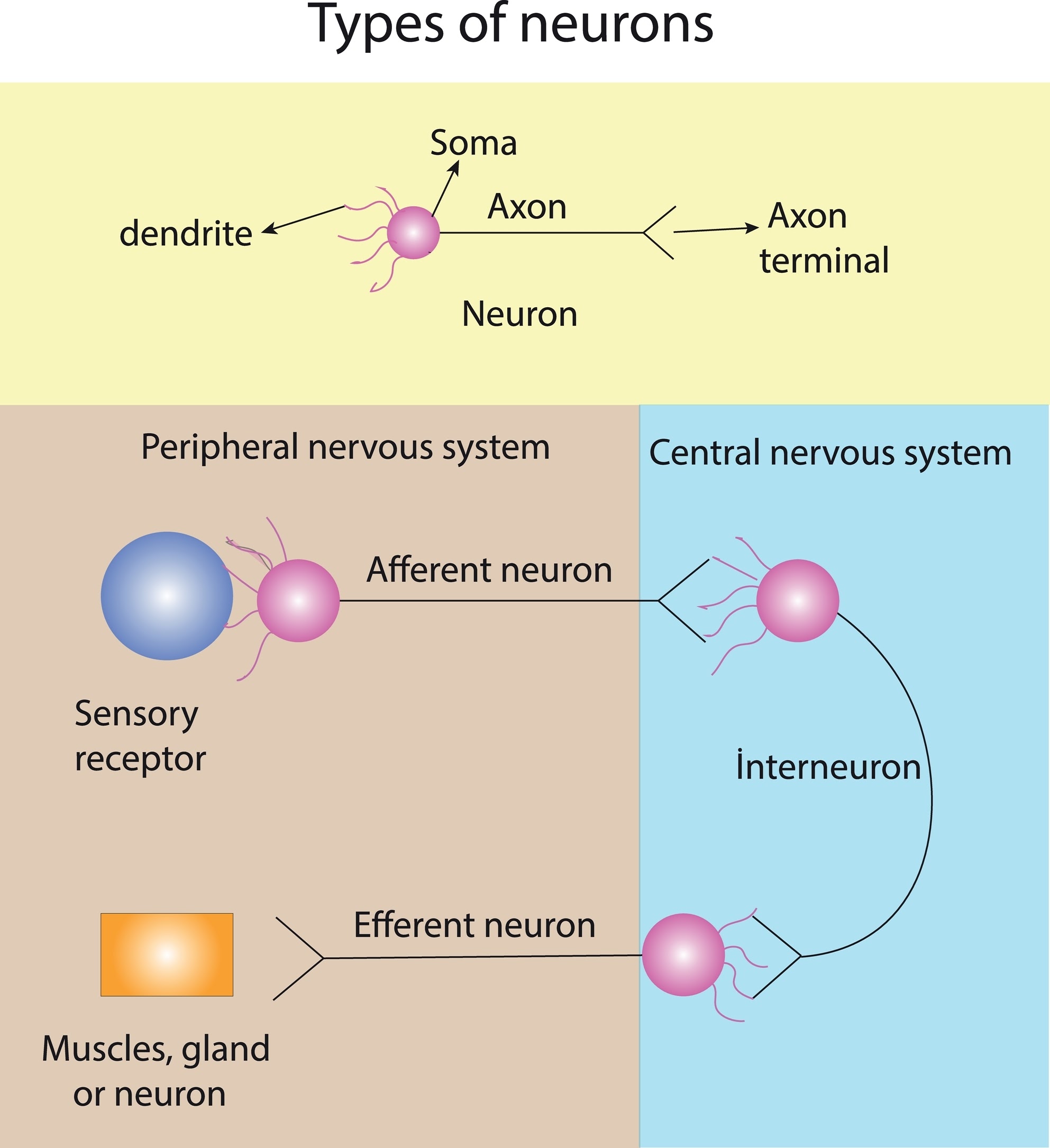
- NeuronsNerve cells; the basic component of the nervous system that provide the means for receiving and sending information through the entire nervous system. are billions of functional units essential for sending and receiving information throughout the nervous system.
- Most neurons share a similar general three-part structure:
- Cell body (soma): Contains the nucleus and organelles, vital for neuron function and homeostasis.
- DendritesExtensions from a neuron’s cell body that receive neural impulses from other neurons; a neuron may have none or as many as thousands of dendrites.: Tree-like branches primarily responsible for receiving signals from other neurons.
- AxonExtensions from a neuron’s cell body that transmit neural impulses to other neurons, structures in the CNS, or muscles; a neuron has only one axon, although most axons branch into many branches.: A long projection that transmits signals away from the cell body to other neurons, muscles, or glands.
- Many axons are covered by a myelin sheathA cellular membrane that speeds up the transmission of neural signals along the axon by acting as an electrical insulator., a fatty layer that acts as an electrical insulator and increases signal speed.
- Axon terminals: Serve as signal transmission relay stations for neurotransmittersChemical signals passed on to other neurons or to muscles in the specific case of movement control., which are chemical messengers.
- SynapseThe junction between the axon of a neuron and another neuron where the passing on of neural signals from one neuron to another occurs.: The specific junction where one neuron communicates with another.
1.3 Neural Communication: The Electrochemical Process
- Neurons communicate through a fascinating electrochemical process.
- When sufficiently stimulated, a neuron generates an electrical impulse called an action potentialA rapid, all-or-nothing electrical signal that travels quickly down the axon once a threshold charge is reached..
- This rapid, all-or-nothing electrical signal travels quickly down the axon once a threshold charge is reached.
- At the axon terminals, the action potential triggers the release of neurotransmitters (chemical messengers stored in vesicles) into the synapse (the tiny gap between neurons).
- Neurotransmitters diffuse across the synapse and bind to specific receptors on the next (post-synaptic) neuron.
- This binding can either excite (making it more likely to fire) or inhibit (making it less likely to fire) the next neuron, ensuring precise information transmission.
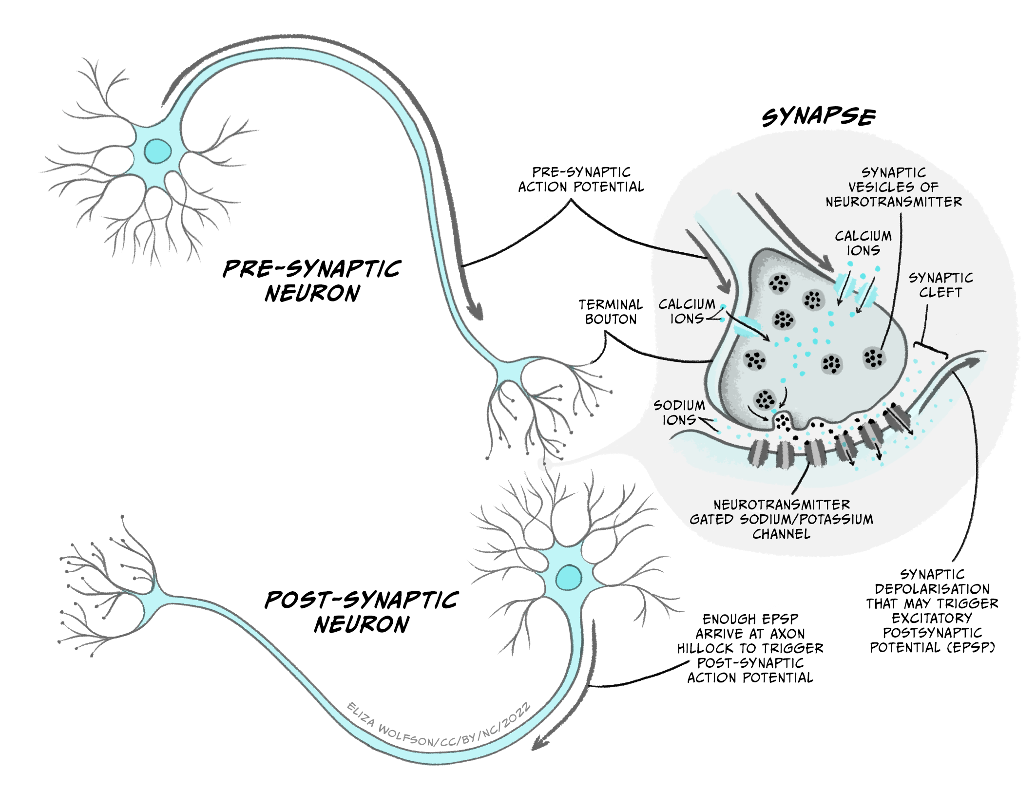
Image credit: https://doi.org/10.20919/ZDGF9829/5
1.4 Functional Classes of Neurons
- There are three main functional classes of neurons based on their role in the Central Nervous System (CNS)The central nervous system functions as the ‘command center’ for human behavior, comprising the brain and spinal cord, and forms the center of activity for the integration and organization of sensory and motor information.:
- 1. Sensory Neurons (Afferent NeuronsNerve cells that send neural impulses to the CNS. They receive information from sensory receptors and convert signals into electrical impulses. They are unipolar with one axon and no dendrites.):
- Send neural impulses to the CNS.
- Receive information from sensory receptors, converting signals into electrical impulses.
- Are unipolarHaving one axon and no dendrites; characteristic structure of sensory neurons. (one axon, no dendrites); cell body mostly in the peripheral nervous system.
- Helpful Tip: Afferent neurons Arrive at the CNS.
- 1. Sensory Neurons (Afferent NeuronsNerve cells that send neural impulses to the CNS. They receive information from sensory receptors and convert signals into electrical impulses. They are unipolar with one axon and no dendrites.):
1.5 Functional Classes of Neurons (continued)
- There are three main functional classes of neurons based on their role in the Central Nervous System (CNS):
- 2. Motor Neurons (Efferent NeuronsNerve cells that send neural impulses from the CNS to skeletal muscle fibers, crucial for movement control.):
- Send neural impulses from the CNS to skeletal muscle fibers, crucial for movement control.
- Alpha motor neuronsMotor neurons found predominantly in the spinal cord that connect directly with skeletal muscle fibers and serve as the functional unit of motor control.: Found mostly in the spinal cord, directly connect to skeletal muscle fibers.
- Gamma motor neurons: Supply intrafusal fibersSpecialized muscle fibers within skeletal muscles that are supplied by gamma motor neurons and are important for muscle spindle function. within skeletal muscles.
- Helpful Tip: Efferent neurons Exit the CNS.
- 2. Motor Neurons (Efferent NeuronsNerve cells that send neural impulses from the CNS to skeletal muscle fibers, crucial for movement control.):
1.6 Functional Classes of Neurons (continued)
- There are three main functional classes of neurons based on their role in the Central Nervous System (CNS):
- 3. InterneuronsSpecialized nerve cells that originate and terminate in the brain or spinal cord; they function between axons descending from the brain and synapse on motor neurons, and between the axons from sensory nerves and the spinal nerves ascending to the brain.:
- Function entirely within the CNS (brain or spinal cord).
- Act as connections between descending axons from the brain to motor neurons, and between sensory nerves and ascending spinal nerves.
- Critical for integrating signals within the CNS.
- Vastly numerous, with an estimated 200,000 interneurons for every sensory neuron.
- 3. InterneuronsSpecialized nerve cells that originate and terminate in the brain or spinal cord; they function between axons descending from the brain and synapse on motor neurons, and between the axons from sensory nerves and the spinal nerves ascending to the brain.:
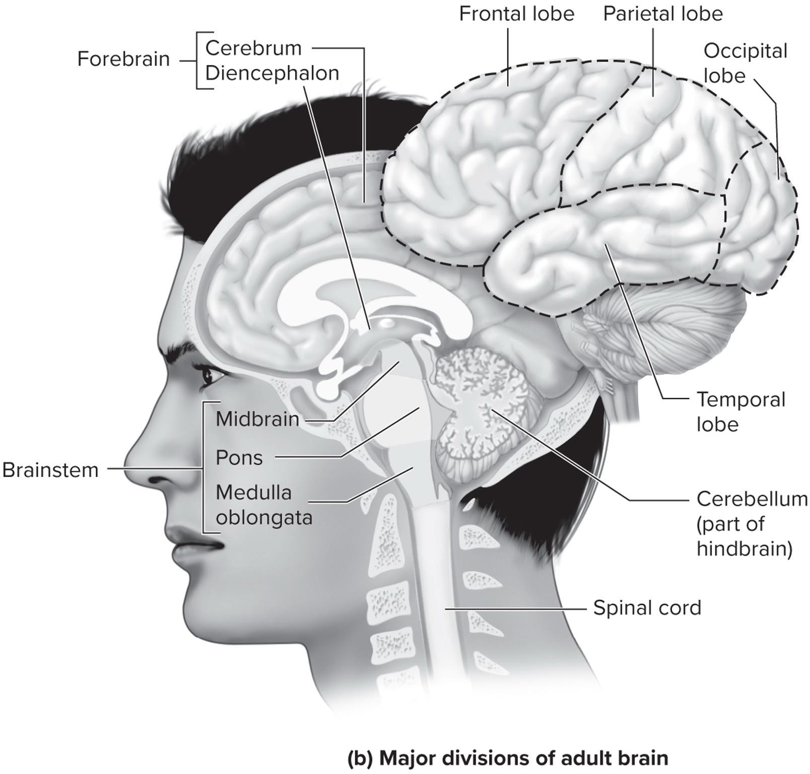
2.2 Major Structural Components of the Brain
- The brain’s major structural components most directly involved in controlling movement are:
- The Cerebrum
- The Diencephalon
- The Cerebellum
- The Brainstem
- The cerebrum and diencephalon are sometimes collectively referred to as the forebrain.
- Figure 4.3 illustrates these major divisions and their subcomponents, helping visualize their location within the brain.
Image credit: (Magill & Anderson, 2016)
2.3 The Cerebrum
- The cerebrumA brain structure in the forebrain that consists of two halves, known as the right and left cerebral hemispheres, connected by the corpus callosum. is divided into two halves: the right and left cerebral hemispheres.
- These hemispheres communicate via the corpus callosumA thick band of nerve fibers that connects the right and left cerebral hemispheres, allowing communication between them., a thick band of nerve fibers.
- The cerebral cortex is the distinctive undulating, wrinkly, gray-colored surface covering both hemispheres.
- It’s a thin tissue (2-5 mm thick) made up of nerve cell bodies, referred to as gray matter.
- Its folding creates gyriRidges formed by the folding of the cerebral cortex that increase its surface area. (ridges) and sulciGrooves formed by the folding of the cerebral cortex that increase its surface area. (grooves), vastly increasing its surface area.
- Cortical neurons are primarily pyramidal cellsPrimary cells for sending neural signals from the cortex to other parts of the CNS, named for the pyramid shape of their cell body., which send neural signals from the cortex to other CNS parts.
- Underneath the gray matter is white matter, an inner layer of myelinated nerve fibers that helps in rapid signal transmission.
Image credit: (Magill & Anderson, 2016)
2.4 Lobes of the Cerebral Cortex
- The cerebral cortex of each hemisphere is divided into four distinct lobes, named for the skull bones closest to them:
- The Frontal Lobe: Located at the front of the brain, anterior to the central sulcus. It is incredibly important for the control of voluntary movement, including planning and executing actions.
- The Parietal Lobe: Situated just behind the central fissure. It is a key brain center for perception and for integrating sensory information from different parts of the body.
- The Occipital Lobe: The most posterior lobe of the cortex. It contains areas vital for visual perception.
- The Temporal Lobe: Located just below the lateral fissure. It plays important roles in memory, abstract thought, and judgment.
Image credit: (Magill & Anderson, 2016)
2.5 Sensory Cortex and Association Areas
- Sensory Cortex Areas:
- Located posterior to the central sulcus, receiving specific types of sensory information via sensory nerves.
- Includes a somatic sensory area processing pain, temperature, and pressure from the body.
- Due to proximity and interconnectedness with motor areas, often referred to as the sensorimotor cortex.
- Association Areas:
- Crucial for higher-level processing, lying adjacent to specific sensory areas (parietal, temporal, occipital lobes).
- They “associate” or connect information from different sensory cortex areas, integrating various types of sensory input.
- Connect with other cortex regions for seamless interaction between perception and higher-order cognitive functions.
- Vital for complex decision-making related to movement, such as in choice reaction time situations.
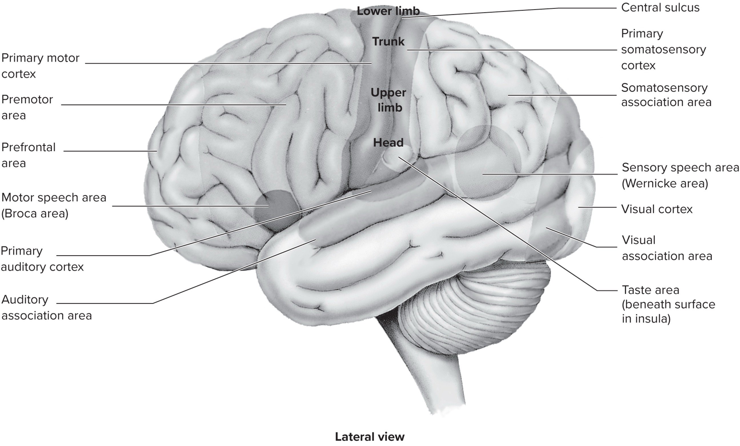
Image credit: (Magill & Anderson, 2016)
2.6 Motor Control Areas of the Cerebral Cortex
- Several areas within the cerebral cortex are particularly active in the control of movement:
- Primary Motor Cortex: Found in the frontal lobe, anterior to the central sulcus. Critical for initiating movements and coordinating fine motor skills (e.g., typing) and postural coordination.
- Premotor Area: Anterior to the primary motor cortex. Essential for organizing movements before they are initiated and maintaining rhythmic coordination during movement. Also plays a role in benefits from observing actions.
- Supplementary Motor Area (SMA): On the medial surface of the frontal lobe. Vital for controlling sequential movements and for the overall preparation and organization of movement.
- Parietal Lobe: A critical cortical area for voluntary movement control . Integrates movement preparation and execution processes by interacting with premotor cortex, primary motor cortex, and SMA before and during movement (e.g., visual attention, grasping).
Image credit: (Magill & Anderson, 2016)
2.7 Brain-Computer Interfaces (BCIs)
- Brain-Computer Interfaces (BCIs) offer incredible potential for individuals with neurological disorders who are unable to move physically .
- BCIs work by reading the electrical activity (brain waves) that occurs in the brain when a person imagines moving a body part .
- They can be part of an EEG skull cap or, in more advanced versions, implanted directly inside the brain .
- With training, BCIs can enable individuals to perform functional activities such as:
- Typing or manipulating small robots .
- Moving a cursor to communicate phrases.
- Maneuvering a prosthetic hand to grasp objects .
- Driving real and simulated wheelchairs .
- The success of BCI research demonstrates its strong potential to benefit a wide range of physical disabilities in the future.
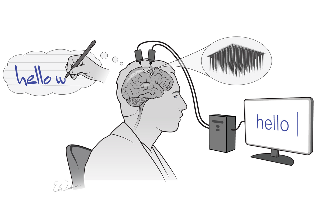
Image credit: https://bit.ly/4fQ9BVK
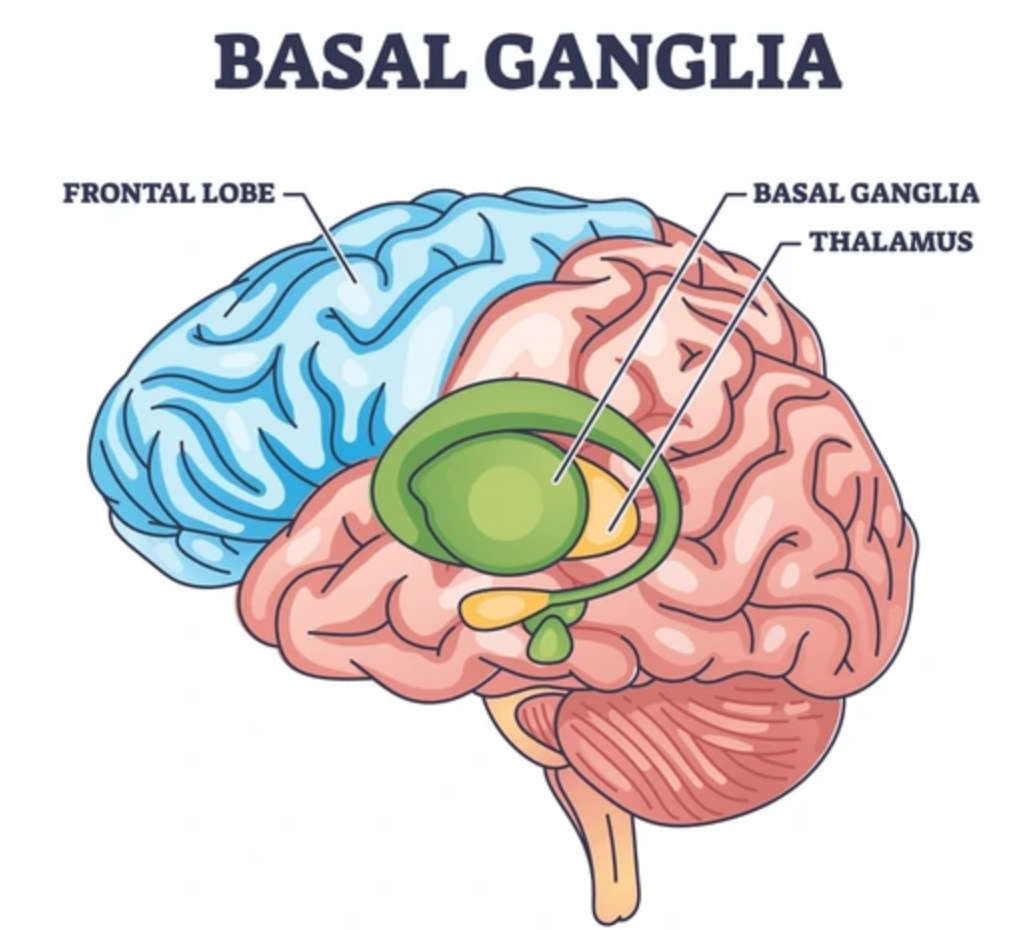
2.8 Subcortical Components: The Basal Ganglia
- The basal ganglia, also known as the basal nuclei, are collections of four large nuclei buried deep within the cerebral hemispheres: the caudate nucleus, the putamen, the substantia nigra, and the globus pallidus.
- They receive neural information from the cerebral cortex and brainstem, with motor neural output primarily to the brainstem.
- The basal ganglia play critical roles in the planning and initiation of movement, the control of antagonist muscles during movement, and the control of force.
- Much is known about their role from studying Parkinson’s diseaseA basal ganglia disorder caused by a lack of dopamine production due to degeneration of neurons in the substantia nigra, leading to bradykinesia (slow movements), akinesia (reduced movement), tremor, and muscular rigidity., a basal ganglia disorder caused by a lack of dopamine production due to degeneration of neurons in the substantia nigra.
- This leads to movement difficulties including bradykinesiaSlow movements, a characteristic symptom of Parkinson’s disease resulting from basal ganglia dysfunction. (slow movements), akinesiaReduced amount of movement, a characteristic symptom of Parkinson’s disease resulting from basal ganglia dysfunction. (reduced movement), tremor, and muscular rigidity.
2.9 Subcortical Components: The Diencephalon
- The diencephalon is strategically located between the cerebrum and the brainstem .
- It contains two important groups of nuclei :
- The ThalamusA crucial relay station that receives and integrates most sensory neural inputs from the spinal cord and brainstem, then passes this information to the cerebral cortex. Also plays a significant role in controlling attention, mood, and pain perception.:
- Acts as a crucial relay station .
- Receives and integrates most sensory neural inputs from the spinal cord and brainstem, then passes this information to the cerebral cortex .
- Also plays a significant role in controlling attention, mood, and pain perception .
- The HypothalamusArguably the most critical brain center for controlling the endocrine system (hormone system), regulating overall body homeostasis including body temperature, hunger, thirst, and physiological responses to stress.:
- Arguably the most critical brain center for controlling the endocrine system (hormone system) .
- Regulates overall body homeostasis, including body temperature, hunger, thirst, and physiological responses to stress.
- The ThalamusA crucial relay station that receives and integrates most sensory neural inputs from the spinal cord and brainstem, then passes this information to the cerebral cortex. Also plays a significant role in controlling attention, mood, and pain perception.:
2.11 The Brainstem
- The brainstem is located directly under the cerebral hemispheres and connects to the spinal cord.
- It contains three main areas involved in motor control :
- Pons: Located at the top, acts as a “bridge” between the cerebral cortex and the cerebellum. Influences chewing, swallowing, salivating, breathing, and balance.
- Medulla (Medulla Oblongata): An extension of the spinal cord. Regulates crucial internal physiological processes like respiration and heartbeat. It is a critical site where sensory and motor corticospinal tracts cross over the body’s midline, meaning one side of the brain controls movements on the opposite side.
- Reticular Formation: A complex network of nuclei and nerve fibers, serving as a vital link between sensory receptors and motor control centers. Its primary role is as an integrator of sensory and motor neural impulses, influencing CNS activity to affect skeletal muscle.
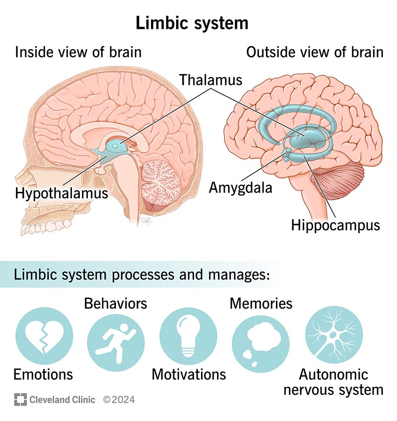
2.12 The Limbic System
- Start by watching this brief video overview of the limbic system: Limbic System Overview.
- The limbic system is not a single structure but an important group of interconnected brain structures.
- It includes parts of the frontal and temporal lobes of the cerebral cortex, as well as the thalamus and hypothalamus, and connecting nerve fibers.
- While often associated with emotions and visceral behaviors, the limbic system also plays important roles in the learning of motor skills.
- This highlights how various brain regions work together to support movement and learning, even those not traditionally thought of as “motor” centers.
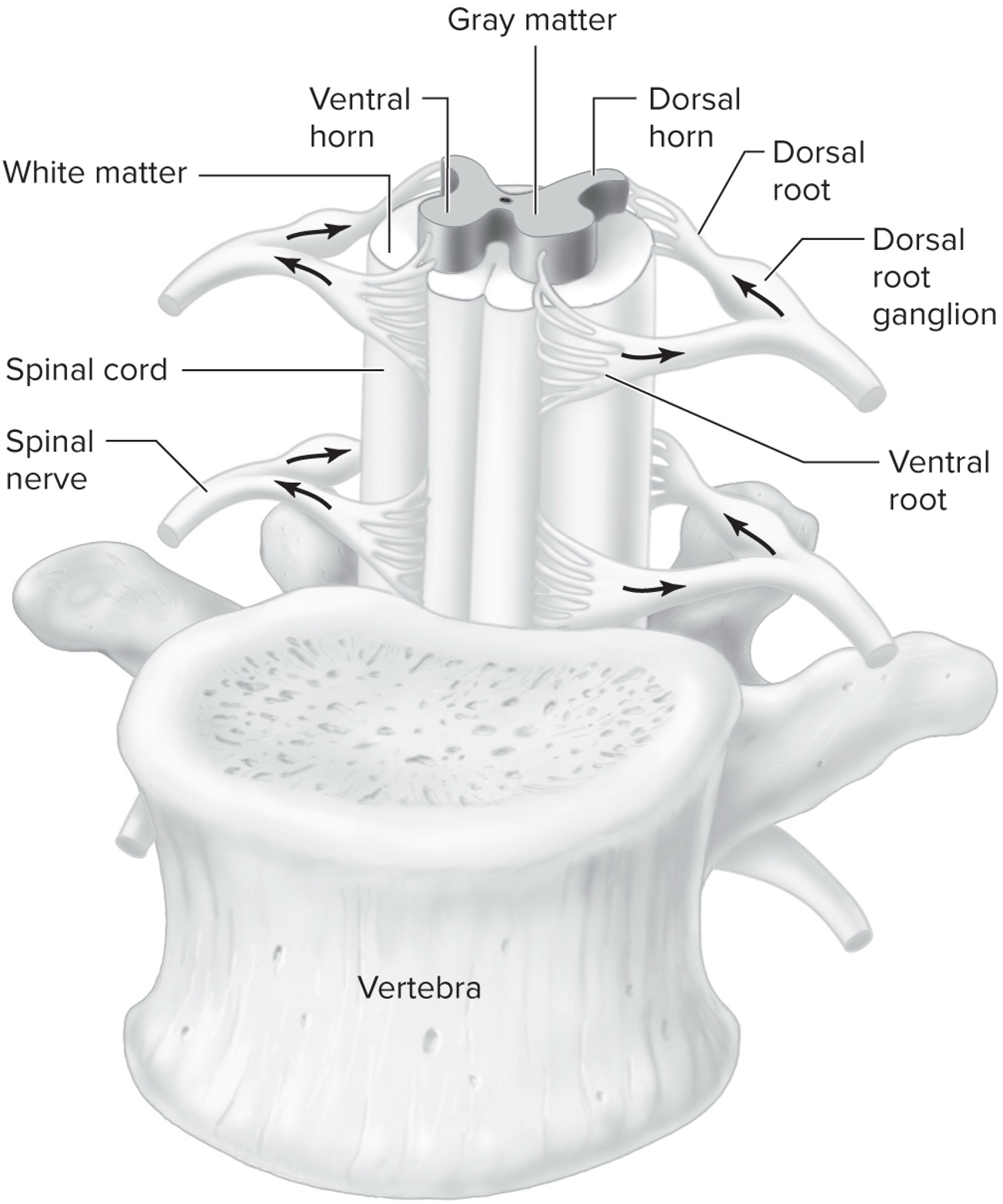
3.1 The Spinal Cord
- The spinal cord is a complex system critically involved in motor control processes and interacts with other systems.
- It is primarily composed of gray matter and white matter.
- The gray matter forms a distinctive butterfly- or H-shape in the central portion:
- Dorsal horns (posterior pair): Contain cells involved in transmitting sensory information from sensory neurons.
- Ventral horns (anterior pair): Contain cell bodies of alpha motor neurons, whose axons terminate on skeletal muscles to initiate movement.
- Also contains interneurons (e.g., Renshaw cells) that can inhibit alpha motor neuron activity.
- The spinal cord also has dorsal and ventral roots where nerves enter and exit, forming spinal nerves.
- It is encased within a vertebra for protection.